What Does a Uterine Fibroid Look Like? Understanding Fibroid Anatomy
Have you ever wondered what’s really going on when you hear the word “fibroids”? If you’re experiencing fibroid-related symptoms like heavy menstrual bleeding, pelvic pain, or even frequent urination, you’re probably looking for clear answers—without all the medical jargon. And if you’ve been diagnosed with uterine fibroids, understanding what they are, how they form, and what they actually look like is an important first step toward feeling empowered about your health.
In this blog, the experts here at VIP Fibroid Center will break down everything you need to know about fibroid anatomy. From the types of uterine fibroids and where they’re found to how they affect your body, we’re here to provide straightforward answers to your most pressing questions.
Whether you’re curious about why fibroids grow, how they can lead to pelvic pressure, or what uterine fibroid treatment options exist, this guide has you covered. By the end, you’ll not only understand what uterine fibroids look like but also feel more confident discussing your symptoms and seeking the care you deserve.
Understanding Uterine Fibroids
Uterine fibroids are incredibly common, but they can feel like a mystery when you’re dealing with symptoms like heavy bleeding, pelvic pain, or even frequent urination. These non-cancerous uterine growths develop for reasons that aren’t entirely clear, but they can impact your daily life in ways that are impossible to ignore. To truly understand what you’re facing, it’s helpful to explore what fibroids are, where they form, and how they might be affecting your body.
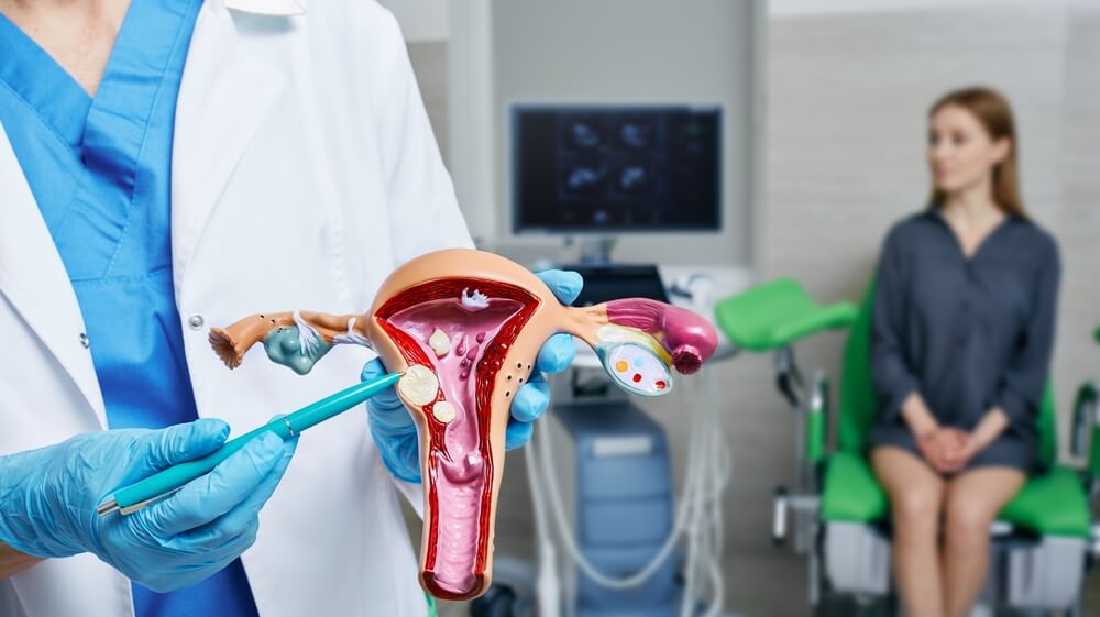
The Anatomy of the Uterus and Fibroids
The uterus is a remarkable organ designed to support life, but it can also be the site of certain conditions like uterine fibroids. Understanding its structure can help you understand how fibroids form and affect your body. Here’s a breakdown:
- Uterine wall: This thick, muscular layer is the powerhouse of the uterus. It stretches during pregnancy and contracts during labor or menstruation, making it a common site for fibroids to develop.
- Uterine cavity: This hollow center of the uterus is where a fertilized egg implants and grows. Fibroids also often form here and cause symptoms like heavy bleeding or menstrual cramps.
- Endometrium: The inner lining of the uterus thickens during the menstrual cycle to support pregnancy. This layer sheds during menstruation, and fibroids near the endometrium can disrupt normal menstrual bleeding.
- Fallopian tubes: These narrow tubes connect the uterus to the ovaries. While fibroids don’t form here, their presence in the uterine cavity can sometimes block these pathways and complicate conception.
- Cervix: This is the lower part of the uterus that opens into the vagina. Fibroids rarely form here but can sometimes push against it, causing discomfort or pelvic pressure.
Fibroids are solid uterine growths made of muscle and connective tissues. Their anatomy varies depending on their type, size, and location in the uterus. Each fibroid type has unique characteristics, but they all share one common trait—they thrive on the blood flow the uterine artery supplies. That’s why treatments like uterine fibroid embolization (UFE) can effectively shrink fibroids by cutting off their supply.
Uterine Fibroids vs. Normal Uterine Tissue
Uterine fibroids differ significantly from normal uterine tissue in both structure and behavior. While the uterus is made of smooth, flexible muscle designed to expand and contract, fibroids are dense, rubbery growths that can range from small and firm to large and irregular. Unlike the uterine wall’s uniform texture, uterine fibroids often feel like nodules or lumps within the otherwise smooth muscle, giving the uterus a distorted shape or size depending on their location.
Normal uterine muscle is soft, pink, and elastic so it can adapt to changes like pregnancy and menstruation. On the other hand, uterine fibroids are usually white or pale blue because of their dense connective tissue and lack of normal blood supply compared to the surrounding uterine tissue. They’re also rigid and unyielding, often causing discomfort or pelvic pressure as they push against surrounding structures.
Their irregularity disrupts the uterus’s natural function, leading to symptoms like heavy menstrual bleeding, frequent urination, and even fertility challenges. Understanding these differences helps explain why fibroids can feel so foreign and disruptive to your body.
Uterine Fibroid Size Ranges
Uterine fibroids can vary dramatically in size, from tiny, barely noticeable nodules to large fibroids that visibly distort the uterus. A fibroid’s size often correlates with the severity of symptoms since larger growths tend to cause more significant pressure and discomfort.
- Small fibroids (<2 centimeters): These fibroids are about the size of a pea or smaller. While they may not cause noticeable symptoms, they can still cause mild menstrual cramps or spotting depending on their location.
- Medium fibroids (2-6 centimeters): Medium fibroids are about the size of a grape to a lemon. They can cause heavy bleeding, frequent urination, or even pelvic pain, especially if they press against the bladder or other organs.
- Large fibroids (>6 centimeters): Comparable to a grapefruit or larger, large uterine fibroids can visibly enlarge the uterus and cause a bloated or protruding abdomen. These fibroids are more likely to result in significant pelvic pressure, prolonged menstrual bleeding, and discomfort during daily activities.
Regardless of size, uterine fibroids can grow over time and shrink with certain treatments like uterine fibroid embolization, which targets the blood flow they rely on to thrive. Identifying the size of your fibroids is a key step in determining the best approach to treating fibroids and relieving your symptoms.
Types of Uterine Fibroids
Fibroids aren’t one-size-fits-all. These uterine growths can vary in where they form, how they behave, and the symptoms they cause. Some grow deep within the uterine wall, while others bulge into the uterine cavity or extend outward. The location and type of fibroid you have can make all the difference in your experience, so it’s important to understand these factors.
Submucosal Fibroids
Submucosal fibroids grow just beneath the uterus lining and extend into the uterine cavity. These fibroids are less common but can significantly impact your symptoms. Because they’re so close to the endometrial lining, submucosal fibroids often lead to symptoms such as:
- Heavy menstrual bleeding
- Prolonged periods
- Menstrual cramps that feel more intense than usual
- Interference with fertility due to distortion of the uterine cavity making implantation difficult
Submucosal fibroids are typically smooth and pale due to their limited blood supply compared to the surrounding uterine tissue. They often protrude into the uterine cavity, creating a bulging or irregular shape that can sometimes be identified during imaging tests. While they’re usually small to medium in size, even a small submucosal fibroid can cause significant symptoms because of its proximity to the endometrium.
Intramural Fibroids
Intramural fibroids are the most common type of fibroid and develop within the uterus’s muscular walls. These fibroids grow deep inside the uterine wall, causing the uterus to feel firm or enlarged. Intramural fibroids can press on nearby organs as they expand, leading to symptoms such as:
- Pelvic pressure
- Frequent urination
- A sensation of fullness in the lower abdomen
- Heavy bleeding
- Menstrual cycle disruptions that can lead to anemia from excessive blood loss
Intramural fibroids tend to start small but can grow to medium or large fibroids depending on hormonal influences. These fibroids may not be as visually obvious as others during imaging because of their location, but they can still distort the uterus’s size and shape.
Subserosal Fibroids
Subserosal fibroids grow on the uterus’s outer surface, just beneath the outermost layer of tissue. Unlike submucosal or intramural fibroids, subserosal fibroids are less likely to cause heavy bleeding since they don’t impact the uterine cavity directly. However, their location can lead to significant pelvic pressure, especially as they grow large enough to push against nearby organs like the bladder or rectum. This can cause symptoms such as:
- Frequent urination
- Constipation
- Lower back pain
Subserosal fibroids often look like rounded bulges on the outer uterine surface. Some are connected directly to the uterus. Others grow on a stalk, making them pedunculated fibroids.
Subserosal fibroids tend to be larger and more irregular in shape, ranging from small, pea-sized growths to masses as large as a grapefruit or even larger in some cases. Their external placement can make them easier to spot during imaging or surgical procedures, though they still pose challenges depending on their size and impact on surrounding structures.
How the Types of Uterine Fibroids Appear During Surgical Procedures
Uterine fibroids are immediately distinguishable from normal uterine tissue during surgical procedures due to their unique texture, color, and placement. Most fibroids have a firm, rubbery feel compared to the uterus’s soft, elastic muscle. Their appearance varies based on their type, size, and location:
- Intramural fibroids: Present as well-defined, rounded masses embedded within the muscle. Surgeons may notice the surrounding tissue stretches and adapts around the fibroid, contributing to an enlarged or irregular uterus shape.
- Submucosal fibroids: May protrude into the uterine cavity, appearing as pale, bulging nodules surrounded by the red, vascular endometrium.
- Subserosal fibroids: Appear as rounded, smooth growth protruding from the outer uterine layer.
- Pedunculated fibroids: Have a mushroom-like appearance with small, firm balls attached by a thin connective tissue band. They can dangle into the uterine cavity or extend outward from the uterus, making them easier to identify and remove.
Regardless of the types of fibroids, their abnormal appearance helps surgeons quickly locate and address them during procedures like UFE or fibroid removal surgery.
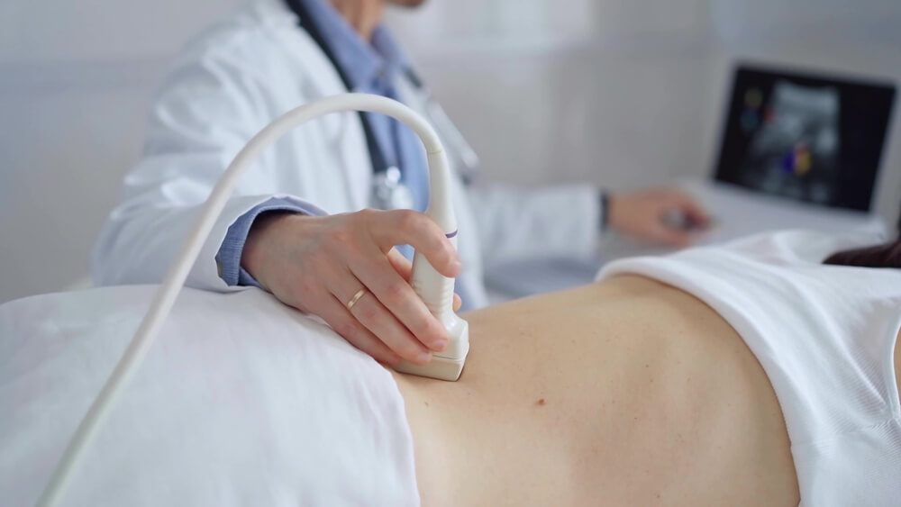
Imaging Techniques to Visualize Fibroids
When you’re dealing with uterine fibroid symptoms, getting a clear diagnosis is key to finding relief. Thankfully, modern imaging techniques can help identify and assess the fibroids to offer insights into their size, type, and location. These tools help you better understand what’s happening inside your body and guide your doctor in creating a treatment plan tailored to your needs. Consider two of the most common imaging techniques.
Ultrasound
Ultrasound is one of the most common and non-invasive imaging techniques used to identify and evaluate uterine fibroids. It’s often the first-line imaging tool because it’s safe, painless, and widely accessible.
Ultrasound uses high-frequency sound waves to create real-time images of the uterus and surrounding structures. During the procedure, a specialist places a transducer on your abdomen (transabdominal) or inserts it into the vagina (transvaginal) to get a closer look at the walls. This is particularly effective for detecting smaller fibroids like submucosal fibroids or identifying irregularities within the uterine wall from intramural fibroids.
MRI
Magnetic resonance imaging (MRI) uses powerful magnets and radio waves to produce highly detailed images of the uterus and surrounding organs. This technique is particularly useful for visualizing large fibroids, assessing their composition, and determining the relationship between fibroids and nearby structures.
Unlike ultrasound, MRI provides a comprehensive view that makes it ideal for complex cases involving multiple or large uterine fibroids. Therefore, MRI is sometimes recommended when other imaging methods don’t provide enough clarity or when precise messaging is needed for advanced treatments.
Take Control of Your Fibroid Health with VIP Fibroid Center
Living with uterine fibroids can feel overwhelming, especially when the related symptoms disrupt your daily life. But understanding what fibroids are, how they form, and the options available for diagnosis and relief is a powerful first step toward reclaiming your comfort and confidence. You don’t have to face this journey alone—there are effective, minimally invasive treatments that can make a real difference.
At VIP Fibroid Center, we’re dedicated to helping you find relief without the need for fibroid surgery or lengthy recovery times. Our skilled and compassionate team specializes in uterine fibroid embolization (UFE), a minimally invasive, proven treatment option to shrink fibroids and improve your quality of life. We take the time to answer your questions, address your questions, and make you feel empowered every step of the way.
Contact VIP Fibroid Center today and let us help you turn your understanding of fibroids toward lasting relief. Together, we’ll create a personalized plan to get you back to feeling your best.