Uterine Fibroid: Imaging, Diagnosis & Treatment Planning
If you’ve been told you may have uterine fibroids, advanced imaging tools help doctors see the uterus clearly, pinpoint the exact location of each fibroid, and create a plan that truly fits your needs. Whether you’re experiencing pelvic pressure, discomfort, or heavy menstrual bleeding, precise imaging gives you and your care team the clarity to move forward with confidence.
Clear answers empower healing. With the right imaging, you can finally understand the cause of your symptoms, work with your doctor to make informed choices about your treatment, and take control of your women’s health – one image at a time.
Imaging Methods for Uterine Fibroids
Accurate imaging is the foundation of every successful uterine fibroid diagnosis and treatment plan. It allows your interventional radiologist to pinpoint the precise location of each fibroid, understand its size and type, and rule out other conditions that may mimic similar symptoms.
These imaging tools reveal how your fibroids affect the uterus, uterine cavity, and surrounding organs. Each of the following imaging modalities plays a unique role in assessing uterine fibroids, so your care team has the information needed to determine the best, most effective minimally invasive procedure for lasting relief.
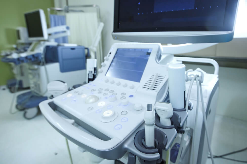
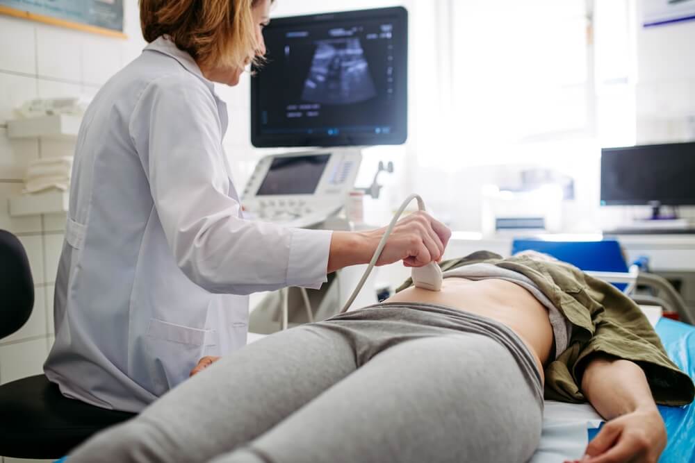
Ultrasound
Often, the first step in identifying fibroids, ultrasound uses safe sound waves to create live images of the uterus and pelvis. It helps detect large fibroids, submucosal fibroids, or calcified fibroids, and shows how they might impact nearby fallopian tubes or the uterine cavity.
Transvaginal Ultrasound
During this specialized form of ultrasound, a small probe is gently placed in the vagina to provide clearer details of the uterine fibroids and their depth within the uterine wall. It’s especially useful for evaluating fibroids causing abnormal bleeding or persistent pelvic discomfort.
Magnetic Resonance Imaging (MRI)
Magnetic resonance imaging (MRI) uses powerful magnets to provide a high-resolution view of the uterus to identify uterine leiomyomas and their exact blood supply. MRI may be helpful in treatment planning for fibroid embolization because it offers insight into smooth muscle cells, tissue composition, and intermediate signal intensity areas that distinguish different fibroid types.
Computed Tomography (CT) Scans
Though less commonly used, CT scans can help visualize calcified fibroids and the overall structure of the pelvis. CT combines multiple X-ray images to show detailed cross-sections of the uterine arteries and surrounding blood vessels, helping rule out rare malignancies or other uterine abnormalities.
The Uterine Fibroid Diagnosis Process
Once imaging is completed, your doctor carefully reviews the results to confirm the presence of uterine fibroids and understand how they may be contributing to your symptoms. The findings help determine the number, size, and position of each fibroid, whether it’s within the uterine cavity, embedded in the wall, or extending outward toward the pelvis.
Your interventional radiologist will also evaluate the blood supply feeding the fibroids. This plays a key role in both fibroid growth and fibroid embolization planning. If you have calcified fibroids, they’ll assess how these hardened areas may affect your overall uterine health and the success of treatment.
In some cases, additional steps such as blood tests or a review of your family history may help identify risk factors that make you more likely to develop uterine leiomyomas. By combining this information with your imaging and reported symptoms, your care team can confirm the diagnosis and build a personalized plan to treat uterine fibroids effectively. This approach ensures that every decision is based on clear evidence and your unique needs as a patient.
Treatment Planning for Uterine Fibroids
After your uterine fibroid diagnosis, your doctor will create a tailored plan based on your imaging results, symptoms, and personal health goals. Factors like the size, number, and location of your uterine fibroids, along with whether they’re calcified fibroids or soft tissue, determine the best course of care. Your interventional radiologist will also consider your age and whether you’ve reached menopause, since fibroids causing pain or abnormal bleeding often respond differently to various treatments.
The goal is always the same—to safely treat symptomatic uterine fibroids, relieve discomfort, and preserve the uterus whenever possible. At this stage, your care team will review all available options. Here are common treatments that may be part of your plan:
- Uterine fibroid embolization (UFE): A minimally invasive procedure where a small catheter delivers small particles to block the uterine artery, cutting off the blood supply to fibroids and causing them to shrink naturally.
- Uterine artery embolization (UAE): Similar to UFE, this treatment targets the uterine arteries feeding the fibroids, reducing abnormal bleeding and pelvic pressure while preserving the uterus.
- Medication therapy: Temporary use of other medicines like gonadotropin-releasing hormone agonists or tranexamic acid may help control bleeding and ease symptoms before definitive treatment.
- Hysterectomy: Surgical removal of the uterus, considered when fibroids are very large or causing severe, recurring problems that don’t respond to other therapies.
- Myomectomy: Surgical removal of individual uterine leiomyomas to preserve fertility; it’s often suggested for younger patients who wish to maintain the ability to become pregnant.
- Lifestyle and dietary management: Adjusting diet, managing stress, and maintaining a healthy weight can help reduce risk factors for new fibroid growth and support long-term women’s health.
Together, these strategies allow your provider to personalize your care. Thus, they can choose the least invasive and most effective approach to help fibroids shrink, improve symptoms, and restore your comfort and confidence.
Take the First Step Toward Clarity and Comfort with VIP Fibroid Center
Understanding what’s happening inside your body brings peace of mind—and the right imaging makes that possible. At VIP Fibroid Center, we combine compassionate care with state-of-the-art imaging and minimally invasive procedures like uterine fibroid embolization. Our experienced interventional radiologists guide you every step of the way so you can get a precise diagnosis and a personalized treatment plan that puts your comfort first.
Contact us today to schedule your consultation and discover how